








1
External Air Supply
You breathe clean air via nasal mask—never inhaling vapor

Biological Dental Treatments
Every infection addressed. Every protocol designed to work with your biology — not against it.
Jawbone Findings
Assessed & Treated
Mercury Fillings
Removed Safely
100% Metal-Free
All Our Materials


IAOMT Accredited Biological Dentist • SMART Certified • SDS Advanced Training • Hürzeler-Zuhr Master Clinician • Dawson Academy Trained • 20+ years of experience







I want them removed safely
Hungary's first SMART-certified clinic — your amalgam fillings release mercury daily.
Those "silver" amalgam fillings are 50% mercury—a known neurotoxin. Every time you chew, brush, or drink hot liquids, they release mercury vapor that your body absorbs.
But there's a safe way to remove them. We're Hungary's first SMART-certified clinic — SMART is the IAOMT's Safe Mercury Amalgam Removal Technique, designed to keep mercury vapor and particles away from you during removal.
Dr. Johanna Malita is SMART-Certified and IAOMT-Accredited.
Conventional removal
Result: mercury vapor and particles reach you
SMART protocol
Result: vapor and particles are kept away from you
You breathe clean air via nasal mask—never inhaling vapor

Seals the tooth completely during removal

Captures mercury vapor at the source, before it reaches the room

Activated charcoal absorbs mercury particles before procedure

Removes filling in large sections—minimizes vapor generation

Reduces vapor release during the entire process
"I want mercury out, but I'm scared removal will make me sicker."
This is a valid concern. Improper removal can expose you to a spike of mercury vapor. That's exactly why SMART protocol was developed—to protect you during removal.
Duration: 60-90 minutes per filling Single-visit for 1-2 fillings Multiple fillings scheduled across sessions
Following IAOMT guidance, we do not remove amalgam during pregnancy or breastfeeding.
Still have amalgam fillings? Send us your X-ray and we will tell you what safe removal involves in your case.
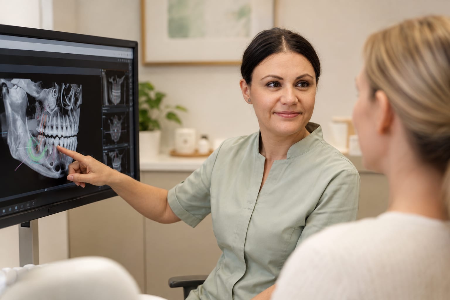
Email your X-rayAfter an extraction the gum can close over the socket while the bone underneath has not fully regenerated. Prof. Shahram Ghanaati calls this a Covered Socket Residuum (CSR). You may have come across the older terms NICO or FDOJ for related jawbone findings — they are not the same diagnosis, and a finding on a scan is not automatically a disease.
Whether it matters is decided with your symptoms, history and examination — never from a picture alone. Some people carry symptoms for years without anyone looking in the jaw.
Surgery that heals faster, hurts less.
What patients notice
Less pain & swelling
Smaller incisions affect less tissue than standard surgery, irritating fewer nerves and blood vessels, so most patients need minimal pain medication.
Faster healing
Precise closures preserve the blood supply, so tissues heal faster. We typically see good healing progress within the first week.
More natural gum contours
Incisions designed to protect the shape of the gum mean tissues heal back to their original form, keeping natural aesthetics for years.
This approach takes more time and specialised training and equipment, but it is designed to protect your tissues and support predictable healing. Dr. Johanna Malita trained with the Hürzeler/Zuhr Academy.

Unwell for years and nobody has looked at your jaw? Send us your X-ray and we will tell you what we can see.
Email your X-ray
Keep it or extract it?
An honest, evidence-based assessment — not a one-size-fits-all answer
Root canal therapy is one of the most debated topics in biological dentistry. Some practitioners remove every root-canal-treated tooth on principle. We don't. Instead, we assess each tooth individually using advanced diagnostics and give you an honest recommendation based on evidence — not ideology.
Where a tooth is infected, medical-grade ozone can be used to disinfect it as part of that assessment — one of the root-canal alternatives we weigh before deciding whether the tooth can be kept.

3D CBCT imaging reveals what standard X-rays miss — hidden fractures, chronic infections at root tips, and bone loss around treated teeth.

We evaluate whether a root-canal-treated tooth is acting as a focal infection — contributing to systemic inflammation, immune burden, or chronic symptoms.

Some root canals can be monitored. Others need retreatment or extraction. We give you the full picture and let you decide — no pressure, no dogma.

Biological extraction with PRF + ozone, followed by a ceramic implant that integrates with your body — not against it.
Unsure whether to keep or replace a root-canalled tooth? Send us your X-ray and we will tell you what your options are.
Email your X-ray
Bleeding gums, bad breath, or a tooth that feels loose?
Biocompatible periodontal care for bleeding gums, deep pockets, and loose teeth.
Gum disease is an infection — not simply a cleaning problem. We identify where the disease is active, treat it carefully, and do everything we reasonably can to help you keep your own teeth.
The elements of our therapy
Healthy gums are not about having your teeth polished more often. We use a combination of modern technology, biological support and practical home care — measured, gentle and tailored to you.


See it. Remove it. Be gentle.
Plaque is not always visible, so we disclose the biofilm first and see exactly where it is accumulating. We then remove it with warm water and fine, low-abrasion AIRFLOW® PLUS powder, using PIEZON® ultrasonics where hardened deposits require them and the PERIOFLOW® nozzle in deeper pockets and around implants. The principle is to clean effectively while avoiding unnecessary trauma to the gums — and as your gum health improves, maintenance can often become gentler.
Clean, support and stimulate healing.
Depending on the area being treated, we may use ozone gas, ozonated treatment water and ozonated oil as adjuncts to professional cleaning. The goal is not only to support local microbial control, but also to create conditions that favour healing of the gum tissues. Ozone does not replace mechanical cleaning; we use it alongside it when we believe it adds value.
When a pocket keeps coming back, we find out why.
If a pocket continues to bleed, remains deep or repeatedly becomes inflamed, we may take a small paper-point sample before cleaning and send it to the IMD laboratory in Berlin, which identifies relevant periodontal bacteria and estimates their quantity. This gives us additional information when a site is not responding as expected, and can help guide a more targeted treatment strategy. Testing can be repeated later to help monitor progress.
Support healing. Reduce discomfort.
Low-level laser therapy, also known as soft-laser therapy, is used for its biostimulatory effect on selected areas after cleaning. It may help reduce discomfort and support the healing response in the gums and surrounding tissues, and can be useful where a site is inflamed, sensitive or prone to bleeding. We use it selectively, when there is a clear reason for it, rather than as a routine add-on.
Because what you do at home makes a real difference.
Periodontal therapy does not end when you leave the treatment room, so our hygienists take the time to teach you exactly how to care for your mouth between appointments: the correct interdental brush size for each space, the right angle and technique, and how to use a water flosser. We show you first on a model, then in your own mouth. You should leave knowing what to do, how to do it, and why it matters.
Look at the bigger picture.
Some periodontal problems are harder to stabilise than others, so we may also discuss factors such as smoking, blood sugar control and nutrition. Where appropriate, we may recommend supportive products or supplements intended to complement professional periodontal care and daily home hygiene — never a replacement for them.
When needed, we go further.
If a pocket remains deep after the first phase of treatment, we reassess the area rather than simply repeating the same cleaning. In selected cases we clean the pocket again and use injectable platelet-rich fibrin (i-PRF), prepared from a small sample of your own blood, with nothing synthetic added. The liquid concentrate is placed locally as an adjunct, to support the healing environment within the treated area.
Keep the progress going.
Where appropriate, we build a home-care routine around your periodontal condition, your anatomy and your response to treatment. It may include ozonated oil, a periodontal toothpaste, a water flosser and interdental brushes — selected and recommended by your hygienist, rather than the same kit for every patient.
More comfortable over time.
By identifying biofilm first and treating only where treatment is needed, we aim to reduce unnecessary trauma to the gums and soft tissues. As periodontal health improves, maintenance treatment can often become gentler and more comfortable.
Our goal is to keep your teeth.
Periodontal therapy is not about making the teeth look cleaner for a few days. The goal is to control the disease, stabilise the gums, and protect the tissues and bone that support your teeth.
“I have had a scale and polish before. It hurt for days, and my gums still bleed. Why would this be any different?”
Because this is not simply another scale and polish. We first identify where disease is active and disclose the biofilm so we can see it, including plaque that is not visible to the naked eye. We then remove it with warm, low-abrasion AIRFLOW® powder and use instruments where deposits actually require them, so that diseased areas are treated thoroughly while healthy tissues are disturbed as little as possible. We cannot promise that periodontal treatment will always be completely discomfort-free. What we can promise is that we measure, explain, work carefully and adapt the treatment to you.
The goal is not simply cleaner teeth today.
It is keeping your gums stable — and keeping your own teeth for as long as possible.
Bleeding gums or a tooth that feels loose? Send us your X-ray and a few words, and we will tell you whether a gum assessment is the right first step.
Email your X-ray
Will my body react to dental materials?
We test how YOUR immune system responds to materials before placement
When we say a material is "biocompatible for most people", we are admitting that some patients develop chronic inflammation from materials their body rejects. Your question should be: "Am I one of them?" Our answer: "Let's find out before we place anything in your mouth."
Your blood is drawn in our office, or a kit is sent to you for collection at your local lab. Simple, quick.
Specialised labs test your blood against 90+ dental materials: composite resins, ceramics, bonding agents, cements and temporaries.
Results show which materials YOUR immune system accepts and which trigger reactions. Green/yellow/red classification. Clear guidance for treatment planning.
We select only materials your body tested green for. No compromises. Only confirmed biocompatible materials for YOUR biology.

Metals and Alloys Sensitivity Screening (Recommended)
Blood test for common problem materials: gold, nickel, palladium, mercury, chromium, cobalt, platinum, copper, silver, tin, plus dental plastics (HEMA, TEGDMA, BISGMA, MMA). Best starting point for most patients.
Titanium Compatibility Evaluation
For patients with existing titanium implants who have complications or unexplained symptoms. Shows whether titanium intolerance may be involved.
Particularly relevant with: autoimmune conditions, chronic inflammation, peri-implantitis, implant failure
Your Exact Materials
Send us the specific products your dentist plans to use - we test those exact materials with your blood. No guessing.
Investment: see the test prices
Timeline: 2-3 weeks for results
Worth noting: Testing beforehand helps you avoid work your body would reject later.
Worried your body may react to a dental material? Send us your X-ray and we will tell you whether testing makes sense for you.
Email your X-ray
I want natural recovery, not just antibiotics
Advanced protocols that support your body's natural healing capacity
Surgery is only half the work. How we prepare for it and support the healing that follows is the other half.
“Here's an antibiotic and a painkiller. Call if problems develop.”
“Your body already has what it needs to heal — we support those natural processes.”
This does not mean we never prescribe antibiotics: where they are genuinely needed, we do. It means we work hard not to over-prescribe them, and this is how we do it.

A small sample of your blood is spun down and the platelet-rich fibrin is placed straight into the surgical site.
Used for: Extractions, implant placement, bone grafting, gum surgery, cavitation treatment.

Medical-grade ozone disinfects the tissue on contact, then converts back to plain oxygen and leaves no residue.
Used for: Extraction sockets, periodontal pockets, implant sites, infected bone.

High-dose intravenous vitamin C supports immune function and collagen formation around a procedure, including after safe amalgam removal.
Used for: Before and after surgery, mercury-removal recovery, longer treatment days.

Low-level light applied to the treated area to support cellular recovery and comfort in the days after surgery.
Used for: Post-surgical healing, swelling and discomfort, soft-tissue recovery.
Detailed pages on each therapy are coming.
FREE · 5 MINUTES · FROM HOME
We'll review your case personally and reply within 3 working days with what we see, what's likely possible, and honest next steps — even if that means we're not the right fit.
What to send
info@johannadental.com
See the full journey — from first email to treatment in Budapest →
No commitment at any stage. You decide if and when to take each step.
Prefer to call? +36-1-701-2900 info@johannadental.com


